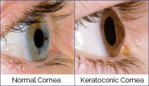
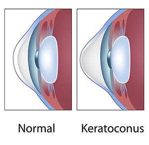
What is Keratoconus?

Keratoconus is a relatively common condition, in which the cornea (the clear front portion of the eye) becomes thin and protrudes. Keratoconus literally means “cone-shaped cornea”. This abnormal shape can cause significant distortion of vision. A keratoconic cornea weakens as collagen fibers deteriorate, causing it to assume a conical shape.

eye diagram

Keratoconus can cause blurred vision, light sensitivity, glare, and decreased night vision. It is usually a progressive condition that worsens over time. In its early stages, it may be treated with prescription glasses or contact lenses.  However, if the condition becomes advanced, cornea transplant surgery may become necessary.

This involves removing the damaged cornea and replacing it with healthy donor tissue. Early detection is critical to preserving a clear vision.  It is recommended those with a family history of keratoconus have annual screenings, especially starting at the age of 12.

Click here to Book Now

What Causes Keratoconus?

Research indicates that keratoconus may be caused by an excess of enzymes that break down the proteins within the corneal surface, causing the cornea to thin and protrude.

It is believed that keratoconus may be genetically inherited. Blood relatives of someone affected with keratoconus may either have a history of keratoconus itself or may have minor changes in their corneas that may indicate that keratoconus probably varies in both specific genetic cause, as well as in its expression within a family. Vigorous eye rubbing can contribute to the progression of the disease. People with keratoconus should avoid rubbing their eyes.

Banner media

normal vs keratoconic cornea

What are the Symptoms of Keratoconus?

Blurred and distorted vision is the earliest symptom of keratoconus. Symptoms usually appear in the late teens or early twenties. The disease will often progress slowly for 10 to 20 years. In the early stages, the vision may only be slightly affected and may progress quickly. Visual blur, distortion, glare, light sensitivity, and corneal irritation are amongst the early signs. Each eye in an individual may be affected differently.

As the disease progresses and the cornea steepens and scars the visual distortion will increase. A sudden visual decrease can occur if the cornea swells. The cornea swells when the elastic part of the cornea develops a tiny crack, created by the strain of the cornea’s protruding cone-like shape. The swelling, known as “corneal hydrops” can persist for weeks to months. As the crack heals it is gradually replaced by scar tissue.

man rubbing his eyes in discomfort

How is Keratoconus Treated?

In order to correct vision, glasses, soft, and “hard” (rigid or scleral) contact lenses are utilized. As the keratoconus progresses, eyeglasses and soft contacts cannot correct the vision sufficiently. When that occurs, only rigid contact lenses can effectively correct vision. When contact lenses cannot improve vision adequately or they become uncomfortable or intolerable, corneal cross-linking (CXL) may be recommended as a means of preventing the need for a corneal transplant. Keratoconus is one of the most common reasons for a cornea transplant.

New Advanced Treatment Now Available to Halt the Progression of Keratoconus – Corneal Cross-linking (CXL)

Corneal cross-linking (CXL), developed as a treatment to halt the progression of keratoconus, has provided excellent results in numerous published studies since it was initially introduced in Europe in 1999.

This minimally invasive procedure uses ultraviolet light which reacts with riboflavin which is applied to the cornea via eye drops. This procedure strengthens the cornea by allowing the cornea to form new “cross-links” between the collagen fibers within the cornea.

This corneal strengthening may not “cure” the disease but evidence suggests that it aids in halting any further progression, and may prevent the need for a corneal transplant in the future. There were only a limited number of sites in the U.S. that were involved in investigational studies of Corneal Cross-linking (CXL) for keratoconus and ectasia. Chicago Cornea Consultants™ was the first practice to offer this procedure in the Chicago area, beginning in 2010, and we have the greatest amount of experience with CXL, performed in over 3000 eyes.

To schedule an appointment for a CXL evaluation, click here to Book Now

Banner media

How Intacs Work

diagram of eyeball

INTACS Corneal Implants

INTACS are revolutionary prescription inserts designed to alter the shape of the cornea for patients with keratoconus. The change in corneal shape causes vision problems that, in early stages, can be corrected by glasses, or, more commonly, rigid contact lenses. In later stages of the disease, a corneal transplant may be required. INTACS can delay or eliminate the need for corneal transplant surgery.

Placed in the periphery of the cornea, INTACS are uniquely designed to help restore the cornea’s natural, dome-like shape. Unlike corneal transplant surgery, INTACS requires no removal of corneal tissue.

NTACS instead provides shape and structure to a cornea affected by keratoconus, helping to maintain or provide a dome-like shape. In certain patients with INTACS who later underwent corneal transplant surgery, their transplants were successfully completed without complications.

To learn more about Intacs click here, or contact Chicago Cornea Consultants™ in Chicago, Highland Park, and Hoffman Estates today.

Would you like to download a brochure about Keratoconus?

Contact us media
Accessibility: If you are vision-impaired or have some other impairment covered by the Americans with Disabilities Act or a similar law, and you wish to discuss potential accommodations related to using this website, please contact our Accessibility Manager at (800) 8-CORNEA.
Contact Us